You have enough on your plate. We make it easy to message your doctor, connect to on-demand virtual care, receive appointment reminders, refill prescriptions, and more.
Healthy for the long haul.
You’re more than a set of symptoms. From pediatrics to adult needs, preventive programs to complex care – we’re here to support you on every step in your journey.
Our doctors do more.
You can enjoy personalized care without pricey membership fees. Our doctors take time to listen to your concerns, from wellness programs to chronic care management.
Proudly part of
Our doctors are based in your community, centered on your needs, and part of a national network that’s transforming healthcare.
Healthcare, handled.
You have enough on your plate. We make it easy to message your doctor, connect to on-demand virtual care, receive appointment reminders, refill prescriptions, and more.
Healthy for the long haul.
You’re more than a set of symptoms. From pediatrics to adult needs, preventive programs to complex care – we’re here to support you on every step in your journey.
Our doctors do more.
You can enjoy personalized care without pricey fees. Our doctors take time to listen to your concerns, from wellness programs to chronic care management.
Compression therapy helps manage swelling, leg ulcer healing rates, decrease the risk of ulcer recurrence and aid the return of blood to the heart.
Compression garments provide external support to the vein walls and work in conjunction with the calf muscle pump.
Medical graduated compression starts at the ankles gradually decreases as it rises up the leg. Compression stockings deliver therapeutic compression for the management of edema,lymphedema and ulcers.
We offer in office sizing with optional styles, colors, sizes, and lengths.
When hardening of the arteries causes a build-up of plaque, a vascular surgeon can first perform angioplasty followed by carotid artery stenting to insert a slender, metal-mesh tube, or stent, to expand the carotid artery to increase blood flow in areas being blocked by plaque. If untreated, enough plaque may build up to reduce blood flow or cause blood clots or pieces of the plaque to break loose and block arteries in the brain.
Carotid artery disease is a serious condition which can cause stroke or arterial embolism, in which a small piece of plaque or a blood clot breaks loose from where it formed and can block another artery further downstream. Clots that block a tiny artery in the brain can cause temporary neurological symptoms, or transient ischemic attacks, also referred to as “mini-strokes.”
For more information on the diagnosis, treatment and procedures regarding vascular disease, visit www.vascularweb.org.
An aneurysm is an enlarged and weakened section of an artery, and is a serious health concern because of the possibility of rupture and blood clots. While they do occur in other arteries, most aneurysms form in the aorta, the largest artery in the human body, running from the heart through the chest and abdomen. The most common type of aneurysms are abdominal aortic aneurysms or AAA.
To surgically repair or remove an aneurysm, the vascular surgeon makes an incision in the skin while the patient is under anesthesia. Through this incision he may perform “open” aneurysm repair or an endovascular procedure called “stent graft” aneurysm repair. The method chosen depends upon location and shape of the aneurysm as well as the health of the patient.
For more information on the diagnosis, treatment and procedures regarding vascular disease, visit www.vascularweb.org.
A safe and long-lasting treatment, carotid endarterectomy is one of the most commonly performed vascular operations. The procedure involves the vascular surgeon removing the lining of the carotid artery that has become damaged or thickened, eliminating plaque from the artery and restoring blood flow.
The carotid arteries are located on each side of the neck and extend from the aorta in the chest to the base of the skull, supplying blood to the brain. Carotid artery disease can create clots on the plaque which can block blood flow to the brain resulting in an ischemic stroke or a mini-stroke. If diagnosed early, treatment can be delivered before a stroke occurs.
For more information on the diagnosis, treatment and procedures regarding vascular disease, visit www.vascularweb.org.
Venous thrombectomy is the surgical removal of a vein clot. This procedure is most commonly used to treat deep vein thrombosis (DVT).
When combined with one or more of the following treatments, venous thrombectomy has a high success rate:
Thrombolysis
Anticoagulant medications
Angioplasty and stenting
Placement of a vena cava filter
When is it indicated?
Venous thrombectomy is indicated in patients with DVT whose symptoms are severe and have been present for fewer than 7 days. Other indications might include:
Severe phlegmasia cerulea dolens
Severe DVT
Patients who should be treated by thrombolysis or anticoagulation therapy
DVT that has not responded to anticoagulation
Patients who are pregnant
For more information on the diagnosis, treatment and procedures regarding vascular disease, visit www.vascularweb.org.
Arteries bringing oxygen-rich blood from your heart to the rest of your body and veins return oxygen-poor blood back to your heart. Varicose veins are swollen veins that you can see through your skin. They often look blue, bulging and twisted. Left untreated, varicose veins may worsen over time.
Venous ablation is a minimally invasive treatment option for varicose veins. It is an outpatient office procedure performed at Vascular Health and Wellness. More than 90% of patients who have undergone venous ablation report improvement or resolution of their leg pain and visibility of varicose veins. Varicose veins can cause aching and feelings of fatigue as well as skin changes like rashes, redness and sores. As many as 40 million Americans, most of them women, have varicose veins.
You have three kinds of veins in your legs: the superficial veins, which lie closest to your skin, the deep veins, which lie in groups of muscles, and perforating veins, which connect the superficial veins to the deep veins. The deep veins lead to the vena cave, your body’s largest vein, which runs directly to your heart. Varicose veins occur in the superficial veins in your legs.
When you are in the upright position, the blood in your leg veins must work against gravity to return to your heart.To accomplish this, your leg muscles squeeze the deep veins of your legs and feet. One-way flaps, called valves, in your veins keep blood flowing in the right direction. When your leg muscles contract, the valves inside your veins open. When your legs relax, the valves close. This prevents blood from flowing in reverse, back down the legs. The entire process of sending blood back to the heart is called the venous pump.
When you walk and your leg muscles squeeze, the venous pump works well. But when you sit or stand, especially for a long time, the blood in your leg veins can pool and the pressure in your veins can increase. Deep veins and perforating veins are usually able to withstand short periods of increased pressures. However, if you are a susceptible individual, your veins can stretch if you repeatedly sit or stand for a long time. This stretching can sometimes weaken the walls of your veins and damage your vein valves. Varicose veins may result. Spider veins are mild varicose veins. They look like a nest of red or blue lines just under your skin. Spider veins are not a serious medical problem, but they can be a cosmetic concern to some people, and they can cause symptoms of aching pain and itching in others.
What are the symptoms?
If you have varicose veins, your legs may feel heavy, tired, restless or achy. Standing or sitting for too long may worsen your symptoms. You may also experience night cramps.
You may notice small clusters of veins in a widening pattern on your leg, or soft, slightly tender knots of veins. Sometimes, the skin on your legs may change color, become irritated, or even form sores.
If you have severe varicose veins, you have slightly increased chances of developing deep vein thrombosis (DVT). DVT may cause sudden, severe leg swelling. DVT is a serious condition that requires immediate medical attention.
What causes varicose veins?
High blood pressure inside your superficial leg veins causes varicose veins. Factors that can increase your risk for varicose veins include having a family history of varicose veins, being overweight, not exercising enough, smoking, standing or sitting for long periods of time, or having DVT. Women are more likely than men to develop varicose veins. Varicose veins usually affect people between the ages of 30 and 70. Pregnant women have an increased risk of developing varicose veins, but the veins often return to normal within 1 year after childbirth. Women who have multiple pregnancies may develop permanent varicose veins.
What tests will I need?
First your physician asks you questions about your general health, medical history and symptoms. In addition, your physician conducts a physical exam. Together these are known as a patient history and exam. Your physician will examine the texture and color of any prominent veins. He or she may apply a tourniquet or direct hand pressure to observe how your veins fill with blood. To confirm a diagnosis of varicose veins, your physician may order a duplex ultrasound test.
Duplex ultrasound uses painless, high-frequency waves higher than human hearing can detect. Your physician uses duplex ultrasound to measure the speed of the blood flow and to see the structure of your leg veins. The test can take approximately 20 minutes for each leg. Besides showing varicose veins, duplex ultrasound may help your physician decide whether your varicose veins could be related to some other condition rather than the veins themselves.
How are varicose veins treated?
Varicose veins may sometimes worsen without treatment. Your physician will first try methods that don’t require surgery to relieve your symptoms. If you have mild to moderate varicose veins, elevating your legs can help reduce leg swelling and relieve other symptoms. Your physician may instruct you to prop your feet up above the level of your heart 3 or 4 times a day for about 15 minutes at a time. When you need to stand for a long period of time, you can flex your legs occasionally to allow the venous pump to keep blood moving toward your heart.
Compression Stockings
For more severe varicose veins, your physician may prescribe compression stockings. Compression stockings are elastic stockings that squeeze your veins and stop excess blood from flowing backward. In this way, compression stockings can also help heal skin sores and prevent them from returning. You may be required to wear compression stockings daily for the rest of your life. For many patients, compression stockings effectively treat varicose veins and may be all that are needed to relieve pain and swelling to prevent future problems.
When these kinds of treatments alone do not relieve your varicose veins, you may require surgical or minimally invasive treatment, depending upon the extent and severity of the varicose veins. These treatments include, ablation and vein stripping.
Ablation
Ablation uses a thin, flexible tube called a catheter inserted into a vein in the leg. Tiny electrodes at the tip of the catheter heat the walls of the vein and destroy it. This mode of treatment frequently replaces the stripping that is performed on the saphenous vein that is described above. The objective is to destroy the saphenous vein that is providing the source for varicose vein development. It can be performed alone or in conjunction with ambulatory phlebectomy, which removes individual clusters of varicose veins from the leg. Your vascular surgeon will advise you regarding which procedure is best for your particular situation.
For more information on the diagnosis, treatment and procedures regarding vascular disease, visit www.vascularweb.org.
Your arteries carry blood rich in oxygen and nutrients from your heart to the rest of your body. When the arteries in your legs become blocked, your legs do not receive enough blood or oxygen, and you may have a condition called peripheral artery disease (PAD), sometimes called leg artery disease.
PAD can cause discomfort or pain when you walk. The pain can occur in your hips, buttocks, thighs, knees, shins or upper feet. Leg artery disease is considered a type of peripheral artery disease because it affects the arteries, blood vessels that carry blood away from your heart to your limbs. You are more likely to develop PAD as you age. One in 3 people age 70 or older has PAD. Smoking or having diabetes increases your chance of developing the disease sooner.
The aorta is the largest artery in your body and it carries blood pumped out of your heart to the rest of your body. Just beneath your belly button in your abdomen, the aorta splits into the two iliac arteries, which carry blood to each leg. When the iliac arteries reach your groin, they split again to become the femoral arteries. Many smaller arteries branch from your femoral arteries to take blood down to your toes.
Your arteries are normally smooth and unobstructed on the inside but, as you age, they can become blocked through a process called atherosclerosis, which means hardening of the arteries. As you age, a sticky substance called plaque can build up in the walls of your arteries. Plaque is made up of cholesterol, calcium and fibrous tissue. As more plaque builds up, your arteries narrow and stiffen. Eventually, enough plaque builds up to reduce blood flow to your leg arteries. When this happens, your leg does not receive the oxygen it needs. Physicians call this leg artery disease. You may feel well and still have leg artery disease or sometimes similar blockages in other arteries, such as those leading to the heart or brain. It is important to treat this disease not only because it may place you at a greater risk for limb loss, but also for having a heart attack or stroke.
What are the symptoms?
You may not feel any symptoms from peripheral artery disease at first. The most common early symptoms is intermittent claudication (IC). IC is discomfort or pain in your legs that happens when you walk and goes away when you rest. You may not always feel pain; instead you may feel a tightness, heaviness, cramping or weakness in your leg with activity. IC often occurs more quickly if you walk uphill or up a flight of stairs. Over time, you may begin to feel IC at shorter walking distances. Only about 50% of the people with leg artery disease have blockages severe enough to experience IC.
Critical limb ischemia is a symptom that you may experience if you have advance peripheral artery disease. This occurs when your legs do not get enough oxygen even when you are resting. With critical limb ischemia, you may experience pain in your feet or in your toes even when you are not walking.
In severe peripheral artery disease, you may develop painful sores on your toes or feet. If the circulation in your leg does not improve, these ulcers can start as dry, gray or black sores and eventually become dead tissue (called gangrene).
What causes PAD?
Athlerosclerosis causes peripheral artery disease. As you get older, your risk of developing leg artery disease increases. People older than age 50 have an increased risk of developing the disease, and men have a greater risk than women.
Other factors that increase your chances of developing the disease include: smoking, diabetes, high blood pressure, high cholesterol or triglycerides, high levels of homocysteine (an amino acid in your blood), and weighing over 30% more than your ideal weight.
What tests will I need?
First your physician asks you questions about your general health, medical history and symptoms. In addition, your physician conducts a physical exam. Together these are known as a patient history and exam. As part of your history and exam, your
physician will ask you if you smoke or have high blood pressure. Your physician will also want to know when your symptoms occur and how often. As part of the physical exam, your physician will conduct pulse tests, which measure the strength of your pulse in arteries behind your knees and feet.
After your exam, if your physician suspects peripheral artery disease, he or she may perform tests, such as:
Ankle-brachial index (ABI), which compares the blood pressure in your arms and legs
Blood tests for cholesterol or other markers for artery disease
To better understand the extent of your leg artery disease, your physician may also recommend duplex ultrasound, pulse volume recording or angiogram
Duplex ultrasound uses high-frequency sound waves to measure real-time blood flow and detects blockages or other abnormalities in the structure of your blood vessels
Pulse volume recording measures the volume of blood at various points in the legs using an arm blood pressure cuff and a Doppler probe
Angiogram, which produces X-ray pictures of the blood vessels in your legs using a contrast dye to highlight your arteries
Physicians usually reserve angiogram for people with more severe forms of leg artery disease
Atherectomy
An atherectomy is a procedure in which your vascular surgeon inserts a specialized catheter into a blocked artery to remove a buildup of atheroclerotic plaque from within the vessel. The catheter contains a sharp rotating blade, grinding bit or laser filament as well as a collection system that permits your surgeon to remove the plaque from the wall of the vessel and collect or suction any resulting debris.
Atherectomy is typically used to treat blockages where angioplasty and stenting cannot be performed. This may be as a result of anatomical factors, the location of the blockage, the hardness of the plaque or other factors. More commonly, atherectomy is uses as a complement to angioplasty and stenting, removing significantly hardened blockages and allowing for the insertion of a balloon and stent. A stent is a small metal device that helps to prevent a blockage from reforming at the same location.
A variety of catheters can be used for this procedure, with the type of catheter being used dependent on the nature of the blockage treated.
For more information on the diagnosis, treatment and procedures regarding vascular disease, visit www.vascularweb.org.
Narrowed blood vessels often need to be expanded to improve blood flow. This can be done with angioplasty, during which your vascular surgeon inflates a small balloon inside the narrowed vessel. Once the vessel is widened, the surgeon may opt to insert a stent, depending on your particular situation. Stents are tiny mesh tubes that reinforce the artery walls to keep the vessels wide open, thus increasing blood flow.
The procedure is typically done through a small incision or puncture in the skin, through which the surgeon inserts a catheter, the tip of which carries the angioplasty balloon or stent. Through x-ray guidance, the surgeon guides the catheter through the blood vessels to the blocked area to insert the balloon of stent. This treatment is most commonly used to treat peripheral arterial disease (PAD), or hardening of the arteries supplying blood to the limbs and organs in the body.
For more information on the diagnosis, treatment, and procedures regarding vascular disease, visit www.vascularweb.org.
This website uses cookies so that we can provide you with the best user experience possible. Cookie information is stored in your browser and performs functions such as recognising you when you return to our website and helping our team to understand which sections of the website you find most interesting and useful.
Strictly Necessary Cookies
Strictly Necessary Cookie should be enabled at all times so that we can save your preferences for cookie settings.
If you disable this cookie, we will not be able to save your preferences. This means that every time you visit this website you will need to enable or disable cookies again.


 When hardening of the arteries causes a build-up of plaque, a vascular surgeon can first perform angioplasty followed by carotid artery stenting to insert a slender, metal-mesh tube, or stent, to expand the carotid artery to increase blood flow in areas being blocked by plaque. If untreated, enough plaque may build up to reduce blood flow or cause blood clots or pieces of the plaque to break loose and block arteries in the brain.
  An aneurysm is an enlarged and weakened section of an artery, and is a serious health concern because of the possibility of rupture and blood clots. While they do occur in other arteries, most aneurysms form in the aorta, the largest artery in the human body, running from the heart through the chest and abdomen. The most common type of aneurysms are abdominal aortic aneurysms or AAA.
 A safe and long-lasting treatment, carotid endarterectomy is one of the most commonly performed vascular operations. The procedure involves the vascular surgeon removing the lining of the carotid artery that has become damaged or thickened, eliminating plaque from the artery and restoring blood flow.
 Venous thrombectomy is the surgical removal of a vein clot. This procedure is most commonly used to treat deep vein thrombosis (DVT).
  Arteries bringing oxygen-rich blood from your heart to the rest of your body and veins return oxygen-poor blood back to your heart. Varicose veins are swollen veins that you can see through your skin. They often look blue, bulging and twisted. Left untreated, varicose veins may worsen over time.
 If you have varicose veins, your legs may feel heavy, tired, restless or achy. Standing or sitting for too long may worsen your symptoms. You may also experience night cramps.
 Varicose veins may sometimes worsen without treatment. Your physician will first try methods that don’t require surgery to relieve your symptoms. If you have mild to moderate varicose veins, elevating your legs can help reduce leg swelling and relieve other symptoms. Your physician may instruct you to prop your feet up above the level of your heart 3 or 4 times a day for about 15 minutes at a time. When you need to stand for a long period of time, you can flex your legs occasionally to allow the venous pump to keep blood moving toward your heart.
 For more severe varicose veins, your physician may prescribe compression stockings. Compression stockings are elastic stockings that squeeze your veins and stop excess blood from flowing backward. In this way, compression stockings can also help heal skin sores and prevent them from returning. You may be required to wear compression stockings daily for the rest of your life. For many patients, compression stockings effectively treat varicose veins and may be all that are needed to relieve pain and swelling to prevent future problems.
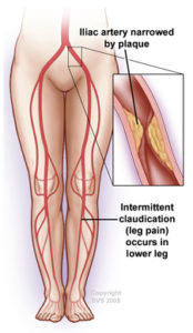 Your arteries carry blood rich in oxygen and nutrients from your heart to the rest of your body. When the arteries in your legs become blocked, your legs do not receive enough blood or oxygen, and you may have a condition called peripheral artery disease (PAD), sometimes called leg artery disease.
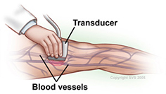 First your physician asks you questions about your general health, medical history and symptoms. In addition, your physician conducts a physical exam. Together these are known as a patient history and exam. As part of your history and exam, your
 Narrowed blood vessels often need to be expanded to improve blood flow. This can be done with angioplasty, during which your vascular surgeon inflates a small balloon inside the narrowed vessel. Once the vessel is widened, the surgeon may opt to insert a stent, depending on your particular situation. Stents are tiny mesh tubes that reinforce the artery walls to keep the vessels wide open, thus increasing blood flow.